Before you travel
Speak to a doctor before your trip
Before you travel, a TreVita physician talks through your history and goals and recommends imaging only when it adds value.
Add-on services · Diagnostic imaging
Imaging helps your physician see exactly what needs attention, so your treatment can be planned with precision. MRI, CT scans, X-rays and ultrasound at accredited partner imaging centers in Baja California, Mexico, 20–30 minutes from San Diego's airport.
Imaging options
4
MRI · CT · X-ray · Ultrasound
Typical scan
20–60 min
Fits into your itinerary
How add-ons work
Your physician recommends it, your coordination team schedules it, and your care team uses the results to plan what comes next.
Before you travel
Before you travel, a TreVita physician talks through your history and goals and recommends imaging only when it adds value.
Before you travel
Your coordination team books the scan around your itinerary. Depending on your case, it may add an extra night to your stay.
In Baja California
Most imaging takes 20 to 60 minutes. You'll arrive knowing where to go, when, and how to prepare.
After your scan
Your physician reviews the results with you and explains what they mean. Whether to move forward with treatment is your decision.
Quick guide
Each type answers a different question. Your physician chooses based on what needs to be seen. Tap a chip to compare.
Soft tissue, joints, spine and brain. The clearest view of cartilage, ligaments, tendons, discs and nerves.
30–60 minutes. Uses magnets and radio waves, so there is no radiation and it is safe to repeat.
Remove all metal and complete a short implant screening. Some scans use contrast; you'll be told in advance.
Bone alignment and joint space. The usual first look at arthritis, and at how a joint behaves under load.
10–15 minutes. Uses a low dose of radiation, so your physician recommends it only when useful.
No fasting or special preparation. Remove jewelry and metal near the area, and tell the team if you could be pregnant.
Detailed bone assessment. Fine bone structure, fracture healing and complex joint anatomy in three dimensions.
10–20 minutes. Uses more radiation than an X-ray, so it is reserved for when the extra detail matters.
Remove metal near the area. Fasting is needed only if contrast is planned; you'll be told in advance.
Tendons and soft tissue, in motion. The lighter option for tendon questions, with no scanner to lie in.
15–30 minutes. Uses sound waves, so there is no radiation and no known risk, including in pregnancy.
No preparation. Wear loose clothing that gives easy access to the area; a water-based gel is used.
Choose an imaging type to see what it's commonly ordered for, who it's for, how to prepare and what the safety screening covers. Your consultation determines which, if any, applies to you.
8 areas
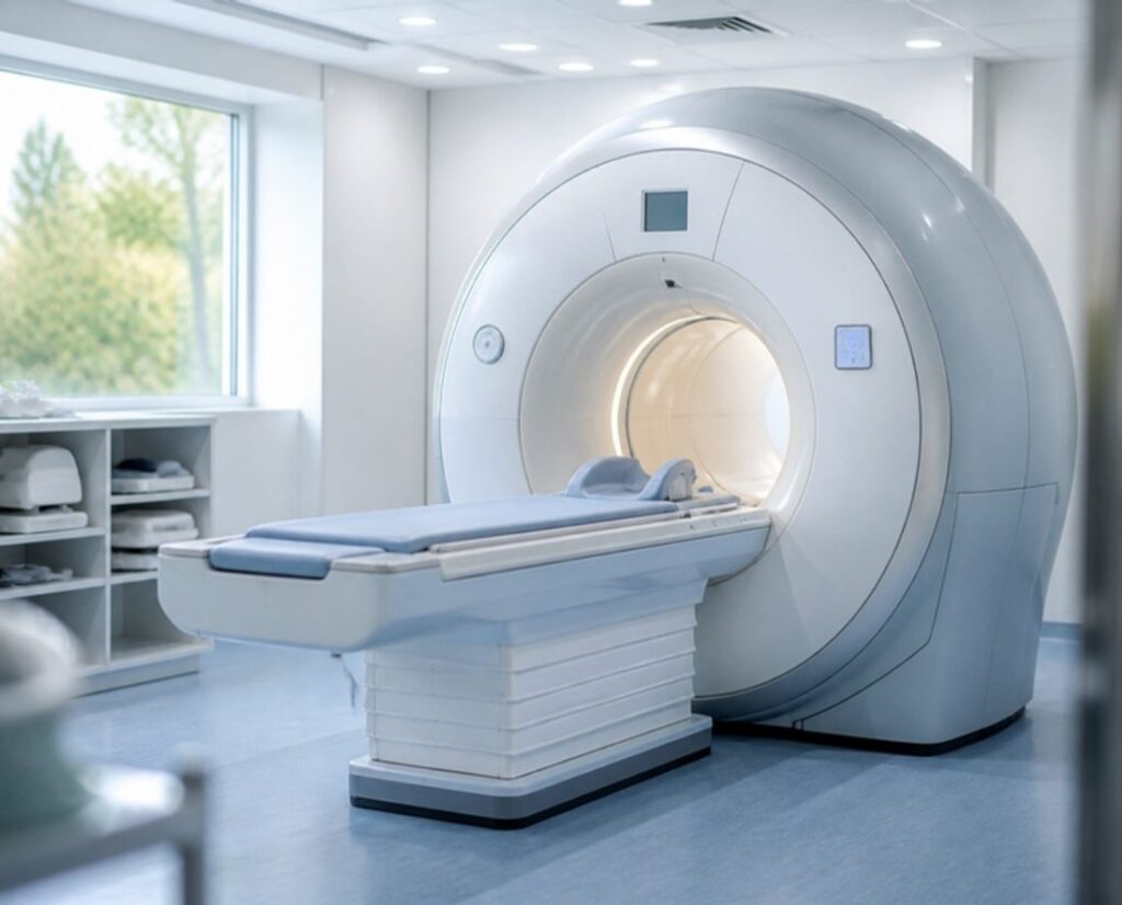
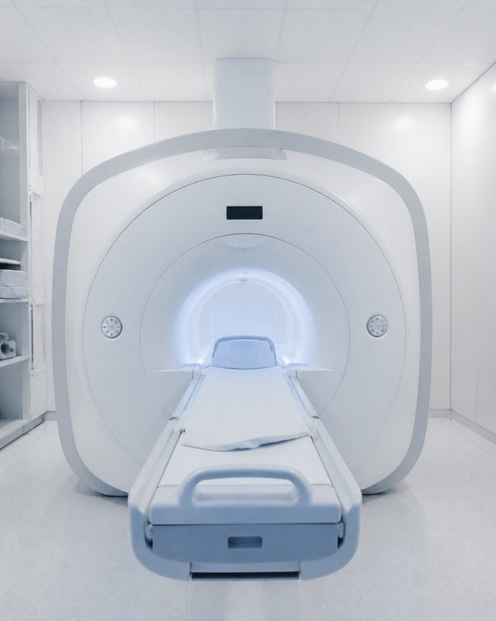
An MRI uses a strong magnet and radio waves to create detailed images with no radiation. It is the clearest way to see cartilage, ligaments, tendons, spinal discs, nerves and the brain. Your physician names the exact study after your consultation.
Grouped by the area scanned. Open a category to see what that MRI is commonly ordered for; your physician names the exact study after your consultation.
An MRI uses a powerful magnet, so you’ll complete a short screening before the scan. Tell your coordinator ahead of time if any of these apply, so the right scan can be planned:
A TreVita physician reviews your particular needs before anything is recommended.
3 study types
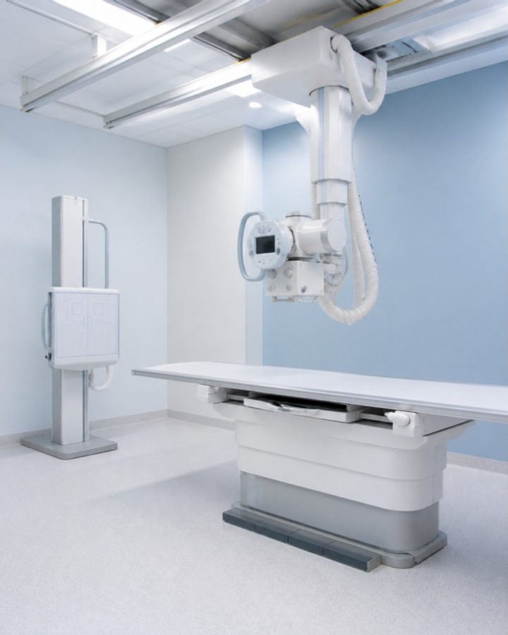
X-rays are the quickest way to see bone: joint alignment, the space left between bones, bone spurs and signs of arthritis. The X-rays recommended through TreVita are taken while you stand or move, so they show how a joint or spine behaves under real load.
Taken while you stand, so the joint is seen under your full body weight. A lying-down X-ray can understate how narrow a joint has become.
A series taken as you bend forward and backward, used to assess whether vertebrae shift more than they should.
Standard images of the affected joints, the usual first look for rheumatoid arthritis, sometimes followed by an MRI.
X-rays use a low dose of radiation, roughly comparable to what you receive from natural background sources over a few days. Tell your coordinator if:
A TreVita physician reviews your particular needs before anything is recommended.
Bone assessment
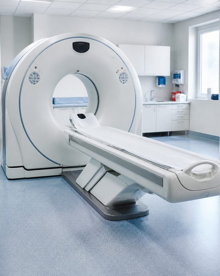
A CT scan combines many X-ray images taken from different angles into detailed cross-sectional and three-dimensional views. For bone, it shows more than a standard X-ray can: the fine structure of a joint, how a fracture is healing, and the shape of complex anatomy such as the pelvis, spine or wrist.
etailed imaging of bone structure and joint surfaces, used to evaluate bone quality, fracture healing, bone spurs and the precise shape of a joint before treatment is planned.
A CT uses more radiation than a plain X-ray, which is why it’s recommended only when the extra detail matters. Tell your coordinator if:
A TreVita physician reviews your particular needs before anything is recommended.
Musculoskeletal
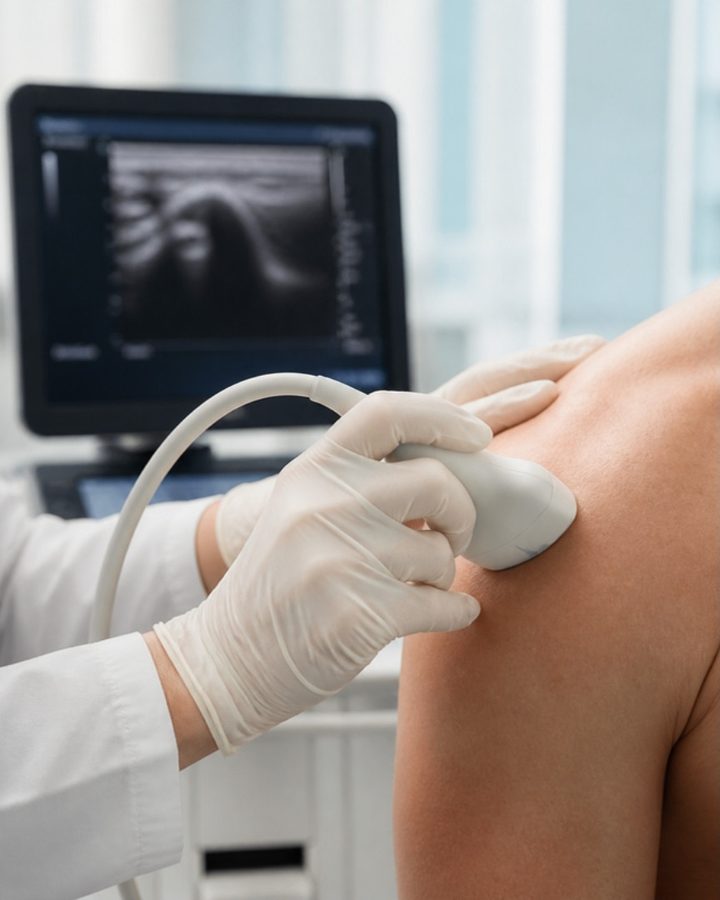
Musculoskeletal ultrasound uses sound waves to produce live images of tendons, ligaments, muscles and the fluid around joints. Because it’s real-time, the specialist can watch a tendon glide as you move, press on the exact spot that hurts, and compare it with the other side. It’s the lighter option for tendon questions; when deeper structures need to be seen, an MRI is the next step.
Targeted imaging of soft tissue and tendons: rotator cuff, Achilles, patellar and elbow tendons, ligaments, muscle tears, bursitis and fluid collections.
Musculoskeletal ultrasound has no radiation and no known risks, and is safe in pregnancy and with implants. The only thing to mention is an open wound or recent surgery over the area, so the exam can be planned around it.

A TreVita physician reviews your particular needs before anything is recommended.
Four ways imaging supports the care you're planning.
Imaging shows your care team the actual condition of the area, so your plan is based on what's there, not on symptoms alone.
Knowing exactly where and how extensive a problem is lets your physician direct treatment precisely, rather than approximately.
A scan taken before treatment gives you a baseline, so progress at follow-up can be seen on an image, not just felt.
Your coordination team fits imaging into your itinerary, 20–30 minutes from San Diego's airport, with or without a procedure.
FAQ's
No. Imaging is recommended by a TreVita physician during your free consultation, and that recommendation is all that’s needed to schedule it.
Yes. Imaging can be scheduled on its own, as part of a procedure trip, or ahead of a decision about treatment. Many clients start with imaging and a consultation before deciding anything else.
Your TreVita physician does, based on your history, symptoms and goals. Each type of imaging answers a different question, and the physician will explain why a particular study was chosen and what it’s expected to show.
Imaging add-ons are quoted individually during your consultation, based on the study your physician recommends. Schedule a free consultation and you’ll receive a clear quote with nothing hidden.
At partner imaging centers in Baja California, Mexico, 20–30 minutes from San Diego’s airport. Your trip is organized and arranged by the coordination team, so the only thing you have to worry about is getting to San Diego.
Often, yes. Share it with your client consultant before your consultation and the physician will tell you whether it’s sufficient or whether an updated or different study would help.
X-rays and ultrasound usually take 10 to 30 minutes. A CT scan takes 10 to 20 minutes. An MRI takes 30 to 60 minutes, longer if contrast is involved. Your coordinator will build the right amount of time into your itinerary.
MRI and ultrasound use no radiation. X-rays use a low dose and CT scans a higher one, which is why your physician recommends them only when the detail they provide matters. Every scan is preceded by a short safety screening, and you’ll always be told what to expect.
It depends on the device. Tell your coordinator before anything is scheduled. In some cases an MRI is still possible; in others your physician will recommend a CT or ultrasound instead.
Let your coordinator know in advance. Shorter scan protocols, positioning, and other options can be discussed with the imaging center so the experience is as comfortable as possible.
Yes. When imaging is part of a procedure trip, it’s typically scheduled early in your stay so the results are in hand before treatment. Your coordinator will confirm the timing in your itinerary.
Your care team reviews the results with you and explains what they mean for your plan, whether that’s confirming treatment, adjusting it, or recommending a different path.
Contact us
A free consultation with the TreVita team is where imaging gets recommended, explained and quoted.